

- HOME
- Research
- Researcher's Profile
- Yoshitaka SHIRASAKI
Researcher's Profile

- Associate Professor
- Yoshitaka SHIRASAKI
- Photonic Imaging
- shirasaki
 g.ecc.u-tokyo.ac.jp
Biography
| April 2004 | Doctoral Research Fellow, Japan Society for the Promotion of Science |
|---|---|
| March 2007 | Doctor of Science, Graduate School of Science and Engineering, Waseda University |
| April 2007 | Project Researcher, Kazusa DNA Res. Inst. |
| April 2009 | SPDR / Researcher, RIKEN RCAI / IMS |
| November 2014 | Project Research Associate, Depertment of Biological Sciences, The University of Tokyo (UTokyo) |
| October 2017 | PRESTO Researcher of Japan Science and Technology |
| April 2019 | Project Research Associate, Depertment of Biological Sciences, UTokyo |
| April 2020 | Project Research Associate, Depertment of Pharmaceutical Sciences, UTokyo |
| August 2023 | Assosicate Professor, RCAST, UTokyo |
Research Interests
We are developing tools for microscopy using the latest optical technologies and microfluidic techniques to understand the biological activities of cells.
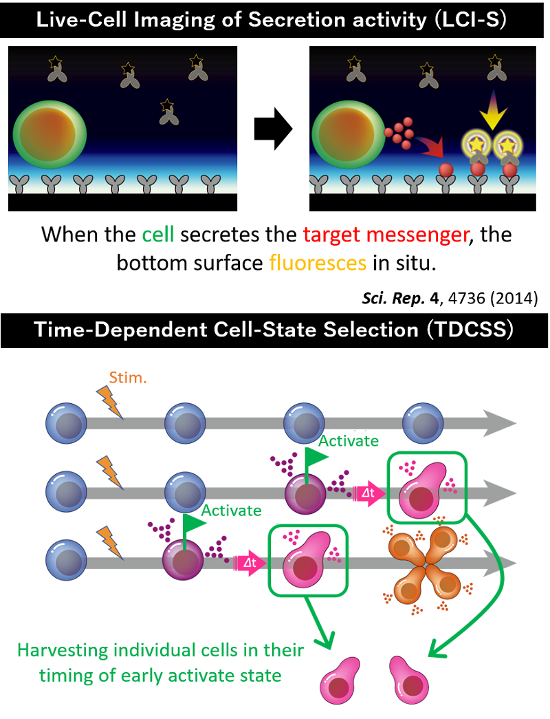
Live-Cell Imaging of Secretion activity (LCI-S): This tool combines an antibody-based sandwich fluorescence immunoassay with total internal reflection fluorescence microscopy to observe cellular secretion in real time (Figure 1 upper).
Time-Dependent Cell-State Selection (TDCSS): This tool tracks cellular changes (e.g., activation or cell death) in real time to find cells that have reached a specific cell-state, harvest them, and examine their gene expression. This allows for a detailed investigation of the genes that control cell activation (Figure 1, lower).
For example, LCI-S visualizes the messages that cells release when they are dying. Cells infected with virus or bacteria cause regulated cell death and release inflammation-associated molecules (Damage-Associated Molecular Patterns: DAMPs) to the extracellular space. However, how exactly DAMPs are released from cells has not yet been elucidated, and LCI-S can simultaneously observe the release of DAMPs and the state of cells, contributing to the elucidation of the mechanism of controlled cell death (Figure 2). LCI-S can also track secretion-associated immune cell activity. Immune cells regulate immune system and maintain the well-being of our bodies by secreting proteins such as cytokines. LCI-S can track the secretion activity of immune cells over several days, enabling us to stratify pathological conditions or TDCSS can investigate genes that control activation of immune cells (Fig. 3).
- Fig.1. LCI-S & TDCSS
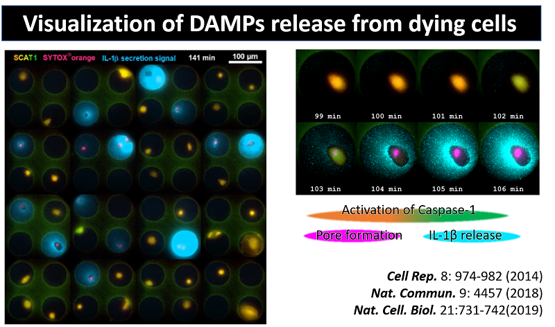
- Fig.2. Visualization of DAMPs release from dying cells
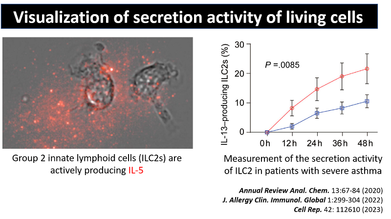
- Fig.3. Visualization of secretion activity of living cells
Award
- October 2015 JAM on cell death 2015, Best Poster Prizes
- September 2019 JSEV2019 Young Investigator Award
Keywords
Optical technologies, Microfluidic techniques, Microscopy
Educational Systems
- Department of Advanced Interdisciplinary Studies, Graduate school of Engineering
