

- HOME
- Research
- Research Category
- Photonic Imaging Ozeki Laboratory
Photonic Imaging
Ozeki Laboratory
Development of advanced photonic imaging methods
Development and application of stimulated Raman scattering (SRS) microscopy
We proposed and demonstrated SRS microscopy, which uses two-color laser pulses to sensitively detect molecular vibrations in biological samples, to visualize biological systems and elucidate the functions of biomolecules by leveraging optical technologies. Furthermore, to improve the molecular discrimination capability of SRS microscopy, we developed a hyperspectral SRS imaging system that acquires SRS images at various molecular vibration frequencies using our original rapid wavelength-tunable laser. We are exploring various biomedical applications, including the analysis of complex structures, dynamics, and interactions within biological systems, through metabolic imaging, super-resolution imaging, and super-multiplexed imaging using Raman-tagged molecules (Figure 1).
Enhancing the sensitivity of SRS microscopy via quantum optics
To realize ultrasensitive SRS microscopy, we are introducing quantum optics (Figure 2), which allows us to break the quantum limit of the signal-to-noise ratio of SRS microscopy. Specifically, by utilizing a special quantum state of light called “squeezed light,” which has smaller quantum fluctuations than classical light, we succeeded in reducing the noise in SRS signals.
Secretion imaging
We have developed "Live-cell imaging of secretion activity: LCI-S," which allows us to visualize the secretion activity of cells in real-time. This technology combines fluorescence sandwich immunostaining and total internal reflection fluorescence microscopy (Figure 3). For example, we can observe the secretion of cytokines, which induce inflammation and allergies, from activated immune cells. We are exploring the medical applications of LCI-S in functional blood diagnostics for precision medicine, phenotypic screening and toxicity assessment for drug discovery, and functional evaluation of cell-based therapeutics.
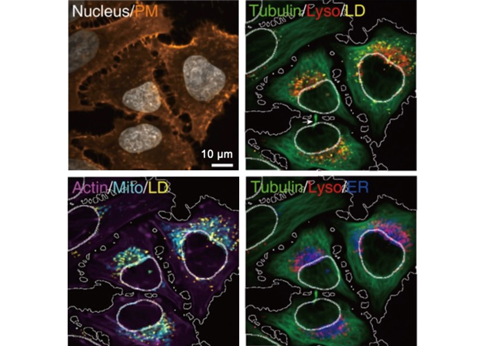
Fig.1. Eight-color imaging of cells with four-color Raman probes and four-color fluorescent probes
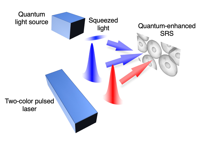
Fig.2. Schematic of quantum-enhanced SRS microscopy
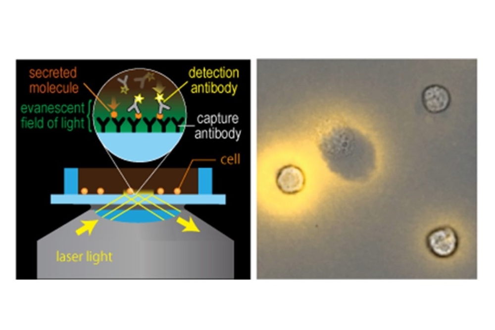
Fig.3. Visualization of cytokine secretion of immune cells by LCI-S
From a young age, my hobbies have been electronics and computers. In university, I majored in electronic engineering and gradually shifted my research focus. Currently, I am advancing research in biophotonics using laser pulses. Understanding the behavior of electronics and optics equips you with tools to develop innovative research through various ingenious methods. Biophotonics is a field that progresses through collaboration with researchers from a wide range of areas, including chemistry and biology. Since we are often unfamiliar with each other's fields, we advance our research by teaching each other the basics, which fulfills my intellectual curiosity every day. My hobby is playing the electric guitar, and the COVID-19 pandemic has given me more opportunities to engage with music.
Member
-
- ProfessorYasuyuki OZEKI
Research Area:Electrical Engineering, Ultrafast lasers, Biophotonics, Raman imaging
-
- Associate ProfessorYoshitaka SHIRASAKI
Research Area:Biophysics, Secretion imaging
Laboratory Homepage
Tags
