

- HOME
- Research
- Researcher's Profile
- Yasuyuki OZEKI
Researcher's Profile
- Professor
- Yasuyuki OZEKI
- Photonic Imaging
- Laser Photonics Sensing
- ozeki
 ee.t.u-tokyo.ac.jp
Biography
| April 2004 | Postdoctoral Researcher of Japan Science and Technology |
|---|---|
| April 2006 | Assistant Professor, Department of Material and Life Science, Osaka University |
| April 2007 | Assistant Professor, Department of Material and Life Science, Osaka University |
| October 2009 | PRESTO Researcher of Japan Science and Technology |
| April 2013 | Associate Professor, Department of Electrical Engineering and Information Systems, The University of Tokyo (UTokyo) |
| June 2021 | Professor, Department of Electrical Engineering and Information Systems, UTokyo |
| April 2023 | Professor, Research Center for Advanced Science and Technology, UTokyo |
Research Interests
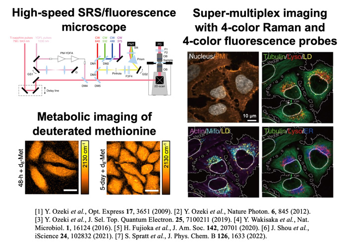
Development and application of stimulated Raman scattering (SRS) microscopy: We proposed and demonstrated SRS microscopy that detects the molecular vibration of biological samples with high sensitivity using two-color laser pulses. Furthermore, to enhance the molecular specificity of SRS microscopy, we developed an SRS imaging system that uses a proprietary high-speed wavelength-tunable laser to obtain SRS images at various molecular vibration frequencies, and are conducting various biological imaging experiments. We are also working on analyzing the complex structure, dynamics, and interactions inside biological samples using Raman probes for metabolic imaging and super-multiplex imaging (Figure 1).
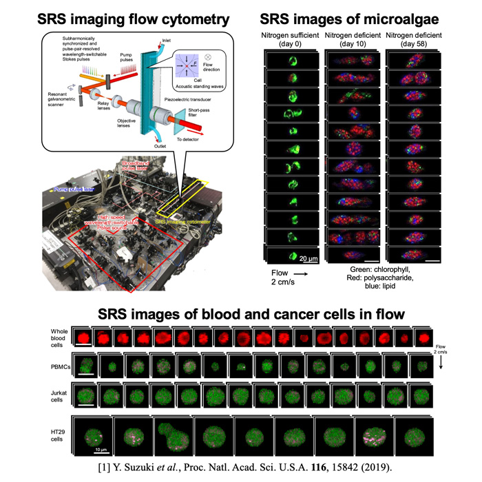
Development and application of large-scale SRS imaging: We have developed an ultra-high-speed wavelength-switchable light source to realize SRS measurements of a large number of cells, and demonstrated multi-color SRS imaging of cells in high-speed flow (Figure 2). This technology makes it possible to visualize biological molecules contained in each of the more than 10,000 cells. We demonstrated the imaging of the nutrients contained in microscopic algae as well as blood cells and cancer cells.
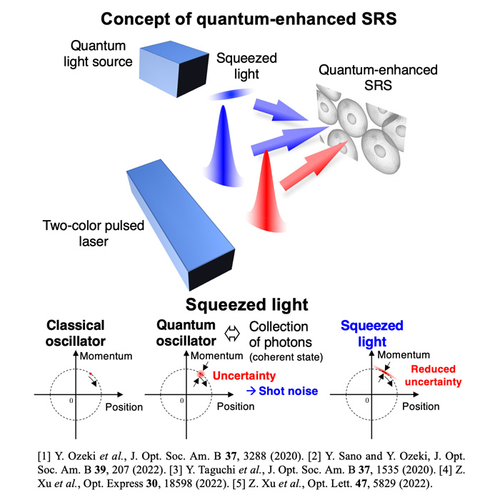
Quantum-enhanced SRS microscopy: We are introducing quantum optics to enhance the sensitivity of SRS microscopy (Figure 3). The signal-to-noise ratio of conventional SRS microscopy is limited by the quantum fluctuations of light. To reduce this fluctuation, we utilize squeezed light to increase the signal-to-noise ratio of the SRS signal.
- Principle of SRS microscopy and its applications
- Large-scale SRS imaging
- Quantum-enhanced SRS imaging
Award
- March 2011 Heisei 22nd Year, Optical Society of Japan Paper Award
- May 2014 Heisei 26th Year, Laser Society Encouragement Award
- February 2017 Heisei 28th Year, Marubun Academic Award.
- February 2020 Reiwa 1st Year, Best Teaching Award, Faculty of Engineering, Utokyo
- April 2020 Reiwa 2nd Year, Minister of Education, Culture, Sports, Science and Technology's Award for Science and Technology
- February 2021 Reiwa 2nd Year, Dean's Award for Research, Graduate School of Engineering, UTokyo
Keywords
Molecular imaging, Ultrashort pulsed laser, Stimulated Raman scattering, Quantum optics
Educational Systems
- Department of Electrical Engineering and Information Systems
- Department of Advanced Interdisciplinary Studies
