

- HOME
- NEWS
- RCAST Report
- The proof of the contribution of mineralocorticoid receptor pathway in blood pressure maintenance during salt restriction
The proof of the contribution of mineralocorticoid receptor pathway in blood pressure maintenance during salt restriction
- Research News
October 12, 2017
Project Researcher Daigoro Hirohama and Emeritus Professor Toshiro Fujita at Research Center for Advanced Science and Technology, the University of Tokyo have demonstrated that the mineralocorticoid receptor (MR)-pendrin system, which plays a role in regulating fluid volume in vivo, contributes to maintaining normal blood pressure during salt restriction.
The paper, entitled "Aldosterone is essential for angiotensin II-induced upregulation of pendrin", has been published in Journal of the American Society of Nephrology on October 11, 2017.
The renin-angiotensin-aldosterone system (RAAS) is known to play an important role in the control of fluid homeostasis and blood pressure (BP) during volume depletion. Dietary salt restriction elevates circulating angiotensin II (AngII) and aldosterone levels, which contributes to maintaining normal BP through the inhibition of salt and fluid loss in the body by increasing NaCl reabsorption in the renal tubules. Up to now, it has been reported that the expression of Cl−/HCO3− exchanger transporter (pendrin) in β-intercalated cells of the renal tubules increases during salt restriction, but the precise mechanism of pendrin activation by RAAS remained unclear. During salt restriction, the mineralocorticoid receptor (MR) which is a receptor of aldosterone is activated, and as a result, various Na+ transporters and Na+ channels are stimulated. Although it has been advocated that MR-pendrin system is involved in the NaCl reabsorption mechanism, its pathophysiological significance remained unclear. Therefore, in this study, we aimed first to elucidate the regulation mechanism of MR-pendrin system by RAAS and further clarify the role of MR-pendrin system in vivo.
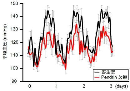
The research team initially administered a low-salt diet to mice and found increased kidney pendrin expression with elevated concentrations of AngII and aldosterone in the plasma. Therefore, in order to remove aldosterone, which is a ligand of MR, from the circulation in mice, analysis was performed using adrenalectomized mice. When AngII alone was administered to adrenalectomized mice, dephosphorylation of MR but not increase in pendrin expression was observed. On the other hand, simultaneous administration of AngII and aldosterone resulted in increased expression of pendrin in addition to MR dephosphorylation. These results suggest that pendrin expression does not increase until aldosterone binds to MR dephosphorylated by AngII. As a result, our results demonstrate that both AngII and aldosterone are indispensable for pendrin activation during salt restriction, and that both have synergistic effects. Next, in order to clarify the in vivo significance of this MR-pendrin system, the effect of dietary salt intake on BP was evaluated in pendrin-knockout mice and wild-type mice. Pendrin-knockout mice showed BP equivalent to that of wild-type mice under a high-salt diet, but showed marked hypotension compared with wild-type mice under a low-salt diet (Fig.). These results reveal that pendrin contributes to maintenance of normal BP during the RAAS activation with a low-salt diet. This suggests that abnormal activation of pendrin develops salt-sensitive hypertension and that pendrin inhibitor can be a new therapeutic agent for salt-sensitive hypertension, and further research in the future will be awaited.
Paper
Aldosterone is essential for angiotensin II-induced upregulation of pendrin
Daigoro Hirohama, Nobuhiro Ayuzawa, Kohei Ueda, Mitsuhiro Nishimoto, Wakako Kawarazaki, Atsushi Watanabe, Tatsuo Shimosawa, Takeshi Marumo, Shigeru Shibata, and Toshiro Fujita
DOI:0.1681/ASN.2017030243
Tags
